How does a DR X-ray machine work to capture and produce digital X-ray images?
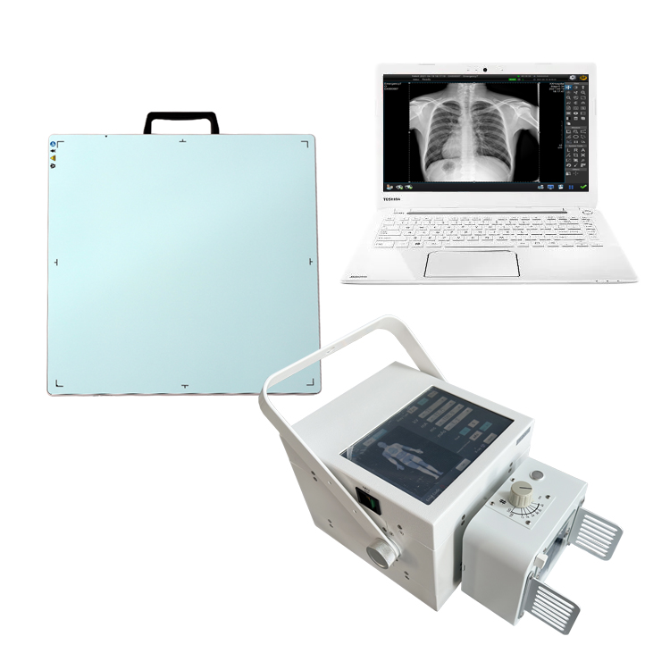
A DR (Digital Radiography) X-ray machine works by using a digital detector to capture X-ray images. Here is a step-by-step explanation of the process:
- X-Ray Generation: The X-ray machine emits X-ray radiation, which passes through the patient’s body or the object being examined.
- Digital Detector: Instead of using traditional X-ray film, a DR X-ray machine employs a digital detector to capture the X-ray image. The digital detector can be either a Flat Panel Detector (FPD) or a Charge-Coupled Device (CCD) sensor.
- X-Ray Conversion: When the X-rays pass through the patient’s body or the object, they interact with the digital detector. The X-rays are converted into an electrical charge pattern on the detector’s surface.
- Analog-to-Digital Conversion: The electrical charge pattern is then converted from analog to digital format. The digital detector divides the charge into small pixels and assigns each pixel a digital value based on the intensity of the electrical charge. This conversion process is known as analog-to-digital conversion.
- Image Processing: The digital X-ray image captured by the detector goes through various image processing techniques to enhance its quality and visibility. These techniques include noise reduction, contrast adjustment, and edge enhancement.
- Image Display: The processed digital X-ray image is displayed on a computer monitor or a dedicated image viewing station. This allows the radiologist or healthcare professional to view the image immediately after it is captured.
- Storage and Distribution: The digital X-ray images can be stored electronically in a Picture Archiving and Communication System (PACS), allowing easy access and retrieval. They can also be shared electronically with other healthcare professionals for consultation or for inclusion in the patient’s electronic medical record.
- Image Analysis and Diagnosis: The radiologist or healthcare professional analyzes the digital X-ray image to make a diagnosis or interpret the findings. They can zoom in, adjust contrast, measure structures, annotate the image, and compare it to previous images, if available.
Overall, the DR X-ray machine replaces the need for film development and enables the immediate production and digital storage of X-ray images, allowing for quicker diagnoses and enhanced workflow efficiency. Whatsapp:+86 18953679166. Email: service@newheek.com