As an important part of the daily work of the imaging department, bedside photography plays an increasingly important role in the examination and diagnosis of critically ill and inactive patients. It not only gains time for the diagnosis and treatment of critically ill patients, but also provides great convenience for patients who are unable to move and are not suitable for activities.
Compared with the mobile CR inspection that was commonly used a few years ago, the image quality of mobile DR is higher than that of mobile CR.
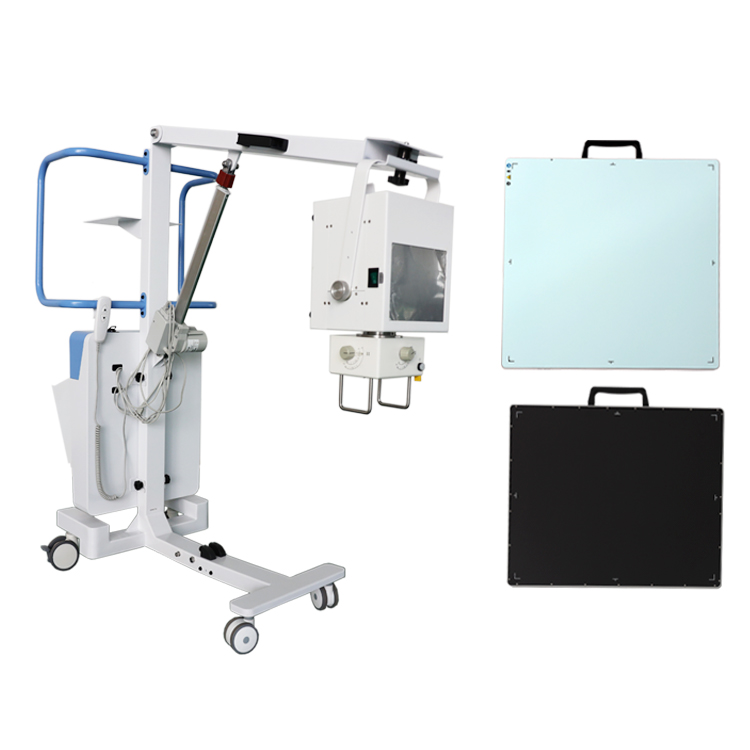
Taking conventional chest photography as an example, we use the same photography conditions to compare mobile DR with a certain brand of CR: although CR has high sensitivity, high spatial resolution, high linearity, large dynamic range and large tolerance And other characteristics, but limited by the density and contrast of the original image, the adjusted image level, clarity and contrast are poor, and the hilar, mediastinum, post-cardiac and subdiaphragmatic structures are not clearly displayed, which may affect the diagnosis; The mobile DR in our hospital uses Canon digital flat panel detectors to collect information for imaging. Its biggest advantage is that it can process the density of the image, and can expand the grayscale image with a resolution of at most dozens of layers to a dynamic range of thousands or even Tens of thousands of layers of gray scale, so it can provide a rich image hierarchy and improve the accuracy of diagnosis of chest diseases.
In addition, mobile DR’s ability to display lesions is also significantly better than mobile CR. Mobile DR is a digital X-ray photography based on a flat panel detector. Therefore, mobile DR has wide dynamic tolerance and large exposure latitude. It can get all the information from skin to bone in a single exposure. Soft tissues all showed good. In addition, the application of equalization technology also shows the advantages of mobile DR post-processing.
In the same image, the thinner or thicker areas of the limbs can be balanced to significantly improve the contrast of the entire area, and at the same time clearly show the anatomical structure of different body thickness areas. This is more practical in the photography of thoracolumbar spine and foot trauma, to avoid missed diagnosis and misdiagnosis. These advantages of mobile DR are lacking in mobile CR.
In addition, mobile DR bedside photography is easy to operate, simple workflow, and fast imaging speed. Digital images can be seen immediately after exposure at the bedside. By adjusting LUT curve, adjusting frequency processing and other post-processing, it can provide high clinical timely Quality imaging for immediate diagnosis.
This will play an important role in responding to emergencies (such as the rescue of earthquake victims). And, because the mobile DR imaging system has a large image storage space (thousands or even tens of thousands of images), it can be taken continuously at the bedside, which makes technicians no longer have to hold a heavy IP board in the radiology and clinical departments. Running back and forth between times greatly reduces the labor intensity of the technicians, and at the same time simplifies the workflow of radiology diagnosis, improving the diagnosis level and work efficiency.
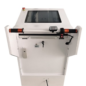
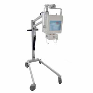
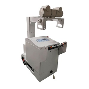
Mobile DR X-ray Machine
Hot Line: +86 18953679166
Whatsapp:+86 18953679166
Email: service@newheek.com
Company: Weifang Newheek Electronic Tech Co., Ltd.
Add: E Building of Future Star Scientific Innovation Industrial Zone of No.957 Wolong East Street, Yulong Community, Xincheng Sub-District Office, Weifang Hi-tech Zone, Shandong Province, China