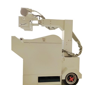
How Bedside Portable DR Addresses Imaging Diagnosis Difficulties of Critically Ill ICU Patients
Traditional Dilemmas in Imaging Diagnosis of Critically Ill ICU Patients
Fatal Transfer Risks: When critically ill patients with numerous life-support devices are transported for examinations, emergencies such as hemodynamic instability, airway dislodgement and sudden oxygenation decline may occur during transit. The incidence of transfer-related adverse events exceeds 30%.
Blind Spots of Static Imaging: Conventional fixed DR only generates single static chest radiographs. It fails to capture functional information including diaphragmatic movement and dynamic changes of pulmonary ventilation, making it difficult to distinguish pulmonary consolidation from transient atelectasis caused by mucus plugs.
Delayed Diagnosis & Treatment: Transfer procedures to and from the radiology department are time-consuming. Immediate screening for critical conditions such as pneumothorax and catheter malposition cannot be realized, which directly impairs the efficiency of clinical decision-making.
Core Advantages of Bedside Portable DR to Break Bottlenecks
Zero Transfer & Bedside Operation:
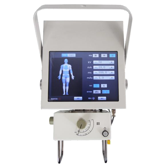
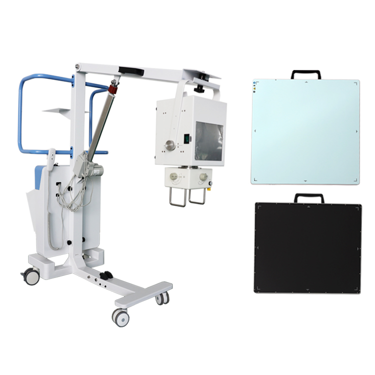
Bedside Portable DR can be directly delivered into ICU wards. Image acquisition is performed without moving patients, completely avoiding secondary injuries and fluctuations in vital signs during transfer. It is hailed as a “mobile imaging center on wheels”.
Upgraded Dynamic Functional Imaging: Advanced models support continuous dynamic image capture lasting several seconds. Clinicians can observe the amplitude, rhythm and coordination of bilateral diaphragmatic motion in real time, accurately evaluating the weaning potential of mechanically ventilated patients and resolving the clinical blind spot that static radiographs cannot detect paradoxical diaphragmatic movement.
High-Definition & Low-Dose Imaging: Equipped with digital flat-panel detectors, bedside Portable DR delivers image quality comparable to fixed-room DR. It clearly displays subtle lung markings and catheter tip positions. Meanwhile, intelligent exposure algorithms reduce radiation dose and minimize unnecessary radiation exposure for both patients and medical staff.
Value in Typical Clinical Scenarios
Precise Catheter Localization: Confirm the positions of endotracheal tubes, central venous catheters and nasoenteric tubes without patient transfer. Visualized guidance during catheter insertion greatly lowers the error rate of blind catheterization.
Rapid Differentiation of Acute Critical Conditions: Imaging observation under dynamic breathing significantly improves the detection rate of supine pneumothorax and free subdiaphragmatic gas, addressing the problem that static radiographs miss over 30% of pneumothorax cases.
Ventilation Function Assessment: Dynamic observation of pulmonary ventilation can be completed at the bedside to distinguish inflammatory consolidation from drainable airway secretions, directly guiding suctioning, postural drainage or adjustment of anti-infection regimens.
Verification via Clinical Practice Case
A patient with weaning difficulty staying in ICU for two weeks only showed blurred shadows at bilateral lung bases on static chest radiographs, which could not be identified as pleural effusion or diaphragmatic weakness. Dynamic imaging from bedside Portable DR clearly revealed nearly absent movement of the left diaphragm and compensatory hyperactivity of the right diaphragm. The core cause — phrenic nerve involvement — was rapidly confirmed, avoiding reintubation caused by premature extubation.
Bedside Portable DR for ICU breaks the spatial restrictions of traditional imaging examinations. It delivers mobile, dynamic and low-radiation imaging services directly to the bedsides of critically ill patients. It fundamentally eliminates transfer risks and fills the gap in functional evaluation existing in static imaging. It has become an indispensable core device within the critical care system.